Uh Oh!
Looks like the cat has chewed the wires again.
No worries! Please click on button to return to the homepage or use the menu above to find what you’re looking for. You may also contact us.
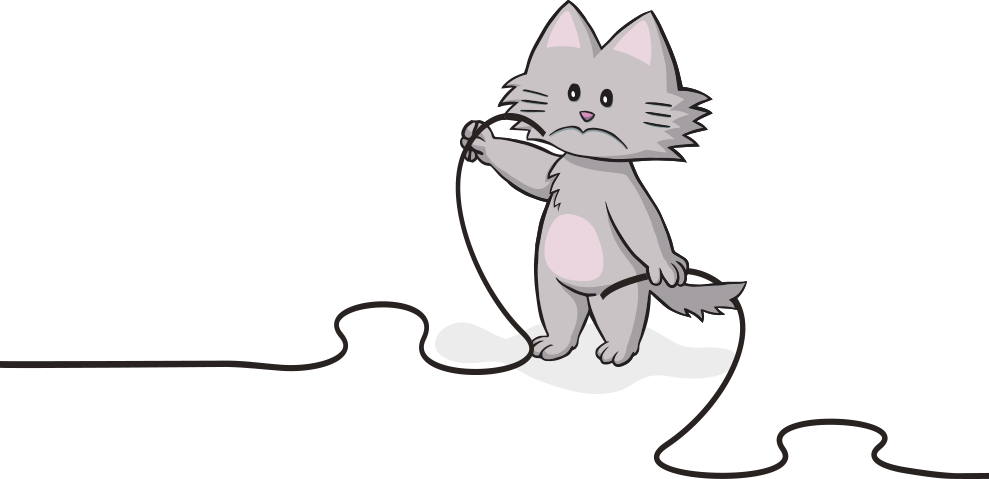

Looks like the cat has chewed the wires again.
No worries! Please click on button to return to the homepage or use the menu above to find what you’re looking for. You may also contact us.
